Drain Removal in Surgical Practice: Timing, Principles and Protocols
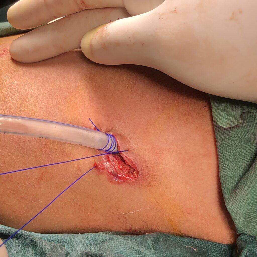
Introduction Surgical drains play a crucial role in postoperative care, helping to evacuate fluids, prevent seroma formation, and monitor complications. However, prolonged drain retention can increase infection risks and tissue irritation. This article outlines evidence-based principles for drain removal, covering timing, techniques, and special considerations across various surgical procedures. When Should a Drain Be Removed? A drain […]
Drain Removal in Surgical Practice: Timing, Principles and Protocols Read Post »