Surgery

How to Present a Radiograph

How to Present a Radiograph: A Step-by-Step Guide

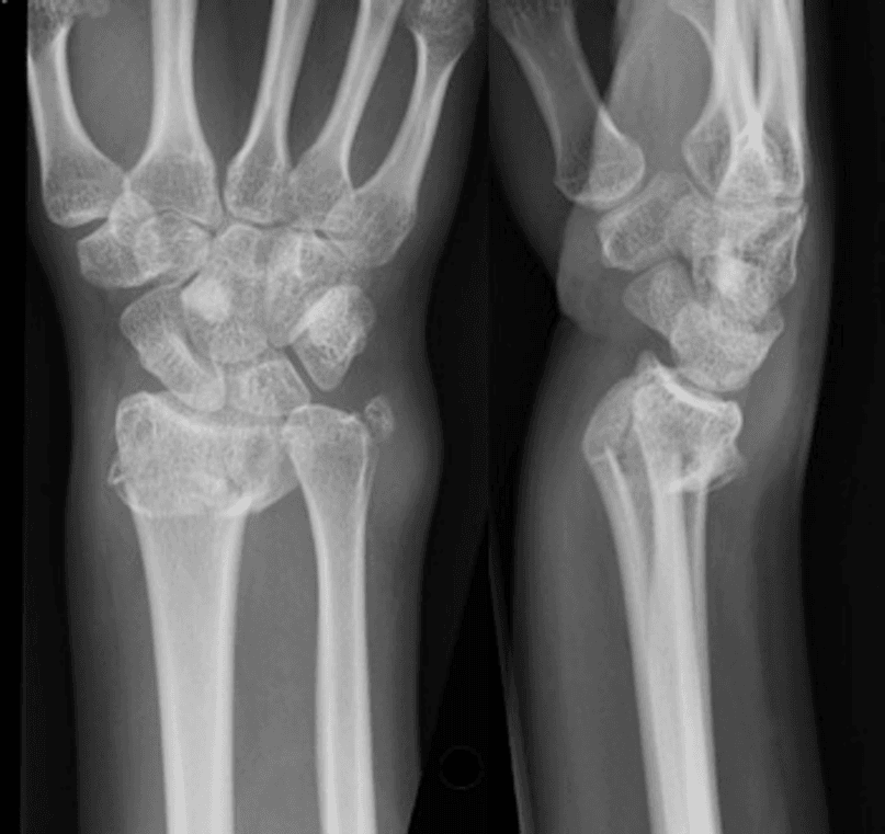
Introduction How to Present a Radiograph? Well, Presenting a radiograph clearly and accurately is essential for effective diagnosis and treatment planning. While classification systems and eponymous terms (like “Colles’ fracture”) can quickly describe fractures, a structured approach ensures thoroughness. This guide will walk you through the key steps to describe a radiograph methodically, covering demographics, […]

How to Present a Radiograph: A Step-by-Step Guide Read Post »

Patient fully recovered with healed and movable arm after fracture trauma – successful bone healing and rehabilitation

Bone Structure And Healing Explained: The 4 Critical Stages

Introduction Bone healing is a remarkable biological process that repairs fractures and restores strength. Whether you’ve suffered a minor crack or a complex break, understanding bone structure and healing phases can help you optimize recovery. This guide covers:✔ Bone anatomy (cortical vs. trabecular bone)✔ Perren’s Strain Theory – why movement affects healing✔ The 4 stages of secondary bone healing✔ Primary vs. secondary healing (and when each occurs)✔ How

Bone Structure And Healing Explained: The 4 Critical Stages Read Post »

Patient with a backslab cast on foot for fracture stabilization – non-weight-bearing orthopedic treatment

Principles of Fracture Management: Reduce, Hold, Move

Introduction Fracture management is a critical aspect of orthopedic care, ensuring patients regain full function with minimal complications. The process revolves around three core principles: reducing the fracture, stabilizing it, and initiating movement for recovery. This guide breaks down essential steps in fracture management, covering reduction techniques, stability assessment, and rehabilitation timing—helping both medical professionals and patients understand the best practices for optimal healing. 1.

Principles of Fracture Management: Reduce, Hold, Move Read Post »

Orthopedic surgeon performing radiological assessment of fractures using digital X-ray display

Radiological Assessment of Fractures – A Complete Guide

Introduction to Radiological Assessment Of Fractures Accurate radiological assessment of fractures is essential for proper fracture diagnosis and treatment planning. This guide covers the key imaging modalities—from basic radiographs to advanced MRI—and explains how to optimize their use in clinical practice. The Golden Rule: Radiograph Fundamentals High-quality radiographs are non-negotiable for proper fracture evaluation. Follow the “Rule of Twos” for

Radiological Assessment of Fractures – A Complete Guide Read Post »

A doctor consults with a patient in a medical facility room, surrounded by healthcare equipment, doing a fracture assessment.

Fracture Assessment: A Complete Guide For Trainees

Introduction to Fracture Assessment Proper fracture assessment is critical for accurate diagnosis and treatment. This guide covers the essential steps—from taking a detailed patient history to performing a thorough physical examination—to help medical professionals identify fractures, assess severity, and determine appropriate management strategies. Step 1: History of Injury Understanding the mechanism of injury is crucial for predicting fracture patterns and associated complications. Key

Fracture Assessment: A Complete Guide For Trainees Read Post »

orthopedic surgeon in a white coat discussing X-ray results on a tablet and illustrating AO fracture classification system.

AO Fracture Classification System

Introduction to Fracture Classification Systems AO fracture classification system help orthopedic surgeons diagnose, plan treatment, and predict outcomes for bone injuries. While many eponymous systems exist for specific fractures, the AO Foundation (Arbeitsgemeinschaft für Osteosynthesefragen) developed a universal, alphanumeric classification system—now widely adopted by groups like the Orthopaedic Trauma Association (OTA). This guide explains the AO fracture classification system, covering diaphyseal (shaft) and metaphyseal (joint-related)

AO Fracture Classification System Read Post »

an orthopedic surgeon in a lab coat holding and examining an X-ray image in a clinical setting, demonstrating fracture types and patterns

ORTHOPAEDIC TERMINOLOGIES – Fracture Types and Patterns

Introduction to Fractures A fracture refers to any break in the continuity of a bone. The term encompasses a wide range of injuries—from severe open, comminuted fractures to subtle undisplaced stress fractures. Fractures are classified based on several key characteristics: Fracture Types and Patterns This guide explores different fracture types, their causes, and clinical significance. Fracture Aetiology: Causes of Bone Breaks

ORTHOPAEDIC TERMINOLOGIES – Fracture Types and Patterns Read Post »

sutures alternatives, surgery, tools, scalpel, scissors, tweezers, vice, needle holder, hegar, paean, needle, operation, procedure, patient, the doctor, medical, hospital, steel, surgery, surgery, surgery, surgery, surgery

Latest Advancement in Sutures Alternatives

Modern Wound Closure: Top Alternatives to Traditional Sutures Introduction to Suture Alternatives While sutures remain a gold standard for wound closure, modern medicine offers several effective alternatives to sutures that provide comparable results with unique advantages. These options are particularly valuable in specific clinical scenarios where sutures may be less ideal. 1. Skin Adhesive Strips: Simple &

Latest Advancement in Sutures Alternatives Read Post »

A healthcare professional carefully suturing a hand, showcasing precision in suture techniques.

Suture Techniques: The 4 Best Methods of Wound Closure

Suturing is a fundamental skill in medical practice, essential for wound closure and optimal healing. Understanding the different suture techniques ensures precision, minimizes scarring, and promotes faster recovery. In this guide, we’ll explore the four most commonly used suturing methods, their applications, and best practices for each. 1. Interrupted Sutures (suture techniques) Interrupted sutures are individually placed and

Suture Techniques: The 4 Best Methods of Wound Closure Read Post »

Close-up image showing a healing scar on a woman's abdomen post-surgery.

Surgical Skin Incisions: Techniques and Principles

Introduction to Surgical skin Incisions Surgical skin incisions: Every surgical procedure begins with a well-planned incision. The way a surgeon makes this initial cut impacts everything from healing time to cosmetic results. This guide covers essential techniques for optimal skin incisions, including blade selection, anatomical considerations, and advanced methods for both open and laparoscopic procedures. The

Surgical Skin Incisions: Techniques and Principles Read Post »

Index
Scroll to Top