Introduction
How to Present a Radiograph? Well, Presenting a radiograph clearly and accurately is essential for effective diagnosis and treatment planning. While classification systems and eponymous terms (like “Colles’ fracture”) can quickly describe fractures, a structured approach ensures thoroughness. This guide will walk you through the key steps to describe a radiograph methodically, covering demographics, fracture assessment (ABCS), and examples of common injuries.
More Ortho Topics: Orthopedics Surgery Series
Step 1: Verify Radiograph Demographics
Before analyzing the fracture, confirm the following details:
- Radiographic view (AP, lateral, oblique, etc.)
- Patient’s name (to avoid misidentification)
- Date of the radiograph (ensures relevance)
- Skeletal maturity (check if growth plates are closed)
Missing any of these could lead to errors in diagnosis or treatment.
Step 2: Assess the Fracture Using the ABCS Method
A systematic approach ensures no critical detail is overlooked. The ABCS framework includes:
A – Adequacy and Alignment
- Is the radiograph of diagnostic quality?
- Are all necessary structures visible?
- Is there proper alignment of bones and joints?
B – Bone Quality and Fracture Configuration
- Assess bone density (osteoporosis, osteopenia).
- Describe the fracture type (transverse, oblique, comminuted).
- Note any displacement, angulation, or shortening.
C – Cartilage (Joint Involvement)
- Is the fracture intra-articular or extra-articular?
- Are there signs of joint subluxation or incongruity?
S – Soft Tissues
- Look for swelling, effusion, or gas shadows.
- Check for foreign bodies or open wounds.
Examples of Radiograph Descriptions
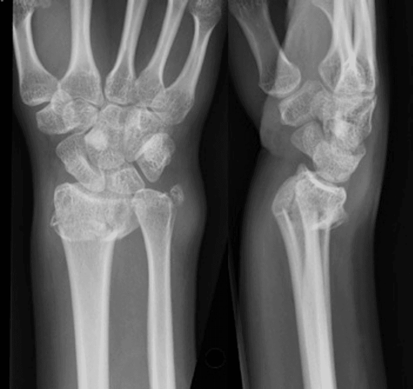
1. Distal Radial Fracture

This radiograph shows AP and lateral views of Mr. Jesse’s left wrist (January 1). The film is adequate, revealing a simple transverse fracture of the distal radius with 20° dorsal tilt and radial height loss. The fracture is extra-articular, with minimal soft tissue swelling.
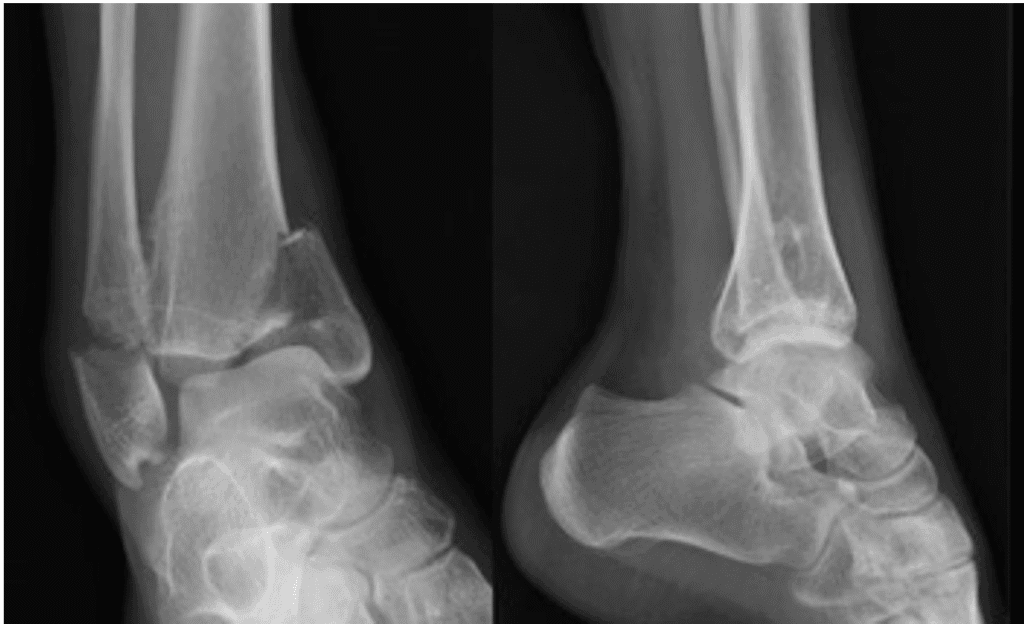
2. Ankle Fracture

This radiograph displays AP and lateral views of Mr. Jesse’s left wrist (January 1). There’s a transverse distal fibula fracture and an oblique medial malleolus fracture with tibial plafond impaction. The ankle joint shows medial subluxation and moderate soft tissue swelling.
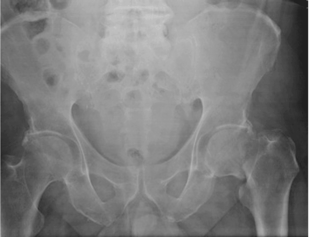
3. Hip Fracture

This is an AP pelvis radiograph of Mr. Jesse’s left wrist (January 1). The right proximal femur is incompletely visualized, but an intracapsular femoral neck fracture with varus angulation is evident. Additional lateral views are needed.
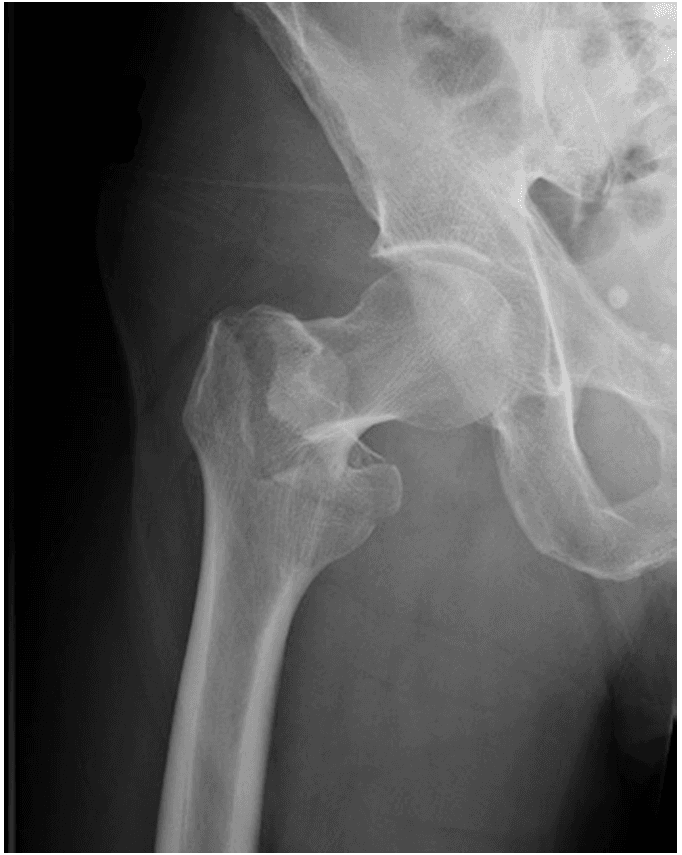
4. Hip Fracture

This an AP hip radiograph of Mr. Jesse’s left wrist (January 1). There’s an intertrochanteric fracture with shortening and varus angulation.
Why Proper Radiograph Presentation Matters
A well-structured description:
✔ Ensures accurate diagnosis
✔ Guides treatment decisions
✔ Improves communication among healthcare providers
✔ Reduces errors in patient management
For further reading on radiographic interpretation, refer to:
Conclusion
Mastering how to present a radiograph improves clinical accuracy and patient care. By following the ABCS method and verifying key details, you can deliver precise and effective fracture descriptions. Whether it’s a wrist, ankle, or hip injury, a structured approach ensures clarity and consistency.
Note: names given are imaginary.
