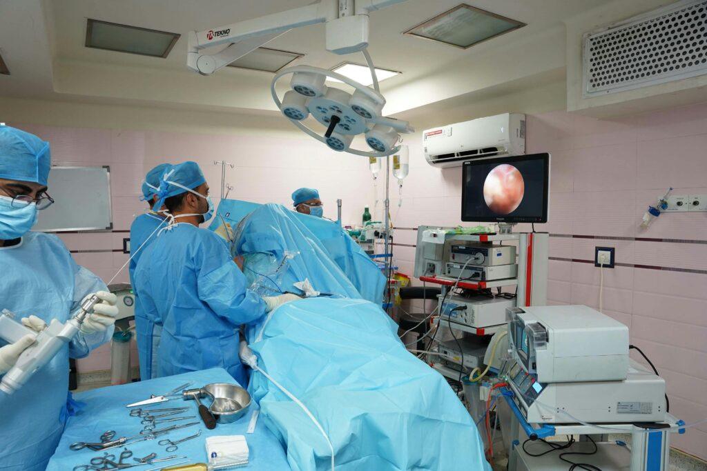
Introduction to Laparoscopic Techniques
Laparoscopic surgery, also known as minimal access surgery, has significantly transformed modern surgical practices. Derived from the Greek words “laparo” (abdomen) and “scopy” (to examine), laparoscopy enables surgeons to examine and operate on the abdominal cavity with minimal incisions. Thanks to advances in optical technology, surgical instruments, and techniques over the past 50 years, laparoscopic surgery has become the preferred choice over traditional open surgery in many procedures.
However, despite its numerous advantages, laparoscopic surgery is not without risks. Studies reveal that many complications arise during the initial access to the abdominal cavity, making safe entry techniques crucial. In this guide, we explore various laparoscopic entry methods, their associated complications, and best practices to minimize risks.

Laparoscopic Entry Methods and Their Conventions – Laparoscopic Techniques
1. Closed (Veress Needle) Method – Laparoscopic Techniques
The Veress needle technique is the most commonly used closed procedure for achieving pneumoperitoneum (CO₂ inflation of the abdomen). The steps involved include:
- A small incision at the umbilicus.
- Insertion of a Veress needle to insufflate the abdomen with CO₂.
- Introduction of a sharp trocar for visualization.
Complications:
- Vascular or visceral injuries: Bowel damage (0.04%) and vascular injury (0.04%).
- Failed pneumoperitoneum due to incorrect needle placement.
- Gas embolism, a rare but life-threatening event.
Safety Concerns:
- Confirm correct needle placement using tests such as the double-click sound, saline drop test, hanging drop test, and aspiration test.
- The most reliable sign is low-pressure insufflation (less than 8 mmHg).
High Intraperitoneal Pressure (HIP) Entry:
- New recommendations suggest starting with 20 to 25 mmHg pressure to reduce the risk of injury by increasing the gap between the abdominal wall and main arteries.
2. Open, Hasson Method – Laparoscopic Techniques
Introduced in 1971 by Harrith Hasson, this method avoids blind access and includes:
- A small fascial incision.
- Insertion of a blunt trocar under direct vision.
- Stabilizing the cannula with stay sutures.
Benefits:
- Zero vascular injury and a significantly lower risk of bowel injury (0.1%).
- Ideal for patients with a history of abdominal surgeries or those who are high weighted.
Problems:
- Slightly higher infection rates (0.4%).
- Increased setup time.
3. Direct Trocar Entry Without Prior Pneumoperitoneum – Laparoscopic Techniques
Developed by Dingerfield in 1978, this technique allows the trocar to be inserted directly without first insufflating the abdomen.
Safety Profile:
- Bowel damage rate: 0.05%.
- Vascular injury rate: 0%.
While faster than the Veress needle approach, it still carries the risk of blind entry, making it less favorable in high-risk patients.
4. Shielded and Optical Trocars (Disposable) – Laparoscopic Techniques
Shielded “safety” trocars were introduced with the idea of preventing contact with intra-abdominal contents. However, studies have shown that shielded trocars are associated with 30% of major laparoscopic entry injuries.
The FDA has cautioned against labeling these devices as “safe” due to insufficient data supporting their claims.
Optical Trocars:
- These devices allow for real-time visualization during entry.
- Although promising, they are still being evaluated to determine if they can surpass conventional techniques in safety and effectiveness.
Laparoscopic Entry Injury Incidence
A review of laparoscopic procedures across various countries highlights the following injury rates:
- Finland: 70,607 procedures, with 1 significant injury per 1,000 procedures (0.6 bowel injuries, 0.3 urological injuries, 0.1 vascular injuries).
- Netherlands: 72 hospitals, 5.7 bowel injuries per 1,000 procedures, with 70% occurring during primary access.
- Australia & UK: 3-6 injuries per 1,000 procedures, with half related to the entry process.
Best Practices for Patient Evaluation and Safe Laparoscopic Entry
Preoperative Evaluation:
- Assess BMI, past surgeries, and potential risk factors (e.g., adhesions or obesity).
- Discuss potential complications, including bowel or vascular injury, and the possibility of converting to open surgery if needed.
Surgeon Expertise:
- Surgeons should be well-trained in various entry techniques and familiar with the instruments and energy sources used during laparoscopic procedures.
Selective Entry Technique:
- For low-risk patients, the direct trocar or Veress needle technique is suitable.
- For high-risk patients (e.g., those with obesity or adhesions), the Palmer’s point entry (left upper quadrant) or the Hasson technique is recommended.
Safety Recommendations:
- Avoid excessive needle movement to prevent enlarging a puncture wound from 1.6mm to 1 cm.
- After entry, always inspect the abdomen for adhesions or injuries.
In Summary: Laparoscopic Techniques
Laparoscopic surgery continues to offer advantages over open surgery in terms of recovery time, pain management, and shorter hospital stays. However, no single entry technique eliminates all risks. The key to safe laparoscopic surgery lies in:
- Proper patient evaluation.
- Surgeon expertise and experience with various entry techniques.
- Adherence to evidence-based guidelines.
As technology advances, newer devices like advanced trocars and optical entry systems may help reduce the risk of complications. For now, surgeon experience and a tailored approach to each patient’s needs remain the most important factors in ensuring success in minimal access surgery.
Additional Reading and Citations
- Royal College of Obstetricians and Gynecologists (RCOG) Policies
- FDA Survey on Shielded Trocars
- Cochrane Review on Laparoscopic Entry Techniques
- Wikipedia: Laparoscopic Approach to Surgery
By staying updated on the latest techniques and best practices, surgeons can continue to improve patient outcomes in minimal access surgery.
