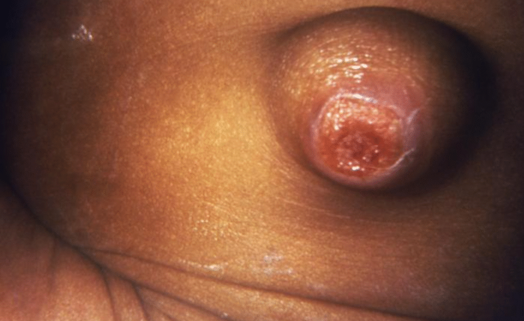
Some children are born with or develop problems that impair the umbilicus, which is an important anatomical feature. It is very important for paediatricians to know the causes, symptoms, diagnosis, and treatment of these disorders. In this section we will read details about umbilical abnormalities.
Read more about paediatric surgery

Acquired Umbilical Abnormalities:
Acquired conditions usually show up after the umbilical cord been cut off from the body.
Umbilical Granuloma
A frequent, non-cancerous growth that happens after the cord is cut. It is marked by a large amount of granulation tissue growing at the base of the umbilical cord.
Pathology:
These lesions are made up of fibroblasts and a lot of capillaries. They range in size from 1 mm to about 1 cm and often have a pedunculated shape.
Management:
- Cauterisation: The first step in treatment is to use silver nitrate to chemically cauterise the area. It may take more than one application for complete epithelialisation to happen.
- Excision: Another option is to surgically remove the tissue and then apply silver nitrate or an absorbable haemostatic substance to the skin.
Things to think about:
Careful use of silver nitrate is very important to avoid burns or skin damage to healthy tissue around the area.
Differential Diagnosis:
If lesions don’t go away after cauterisation, they could be a real umbilical polyp or a sinus tract that won’t go away.
Umbilical Infections (Omphalitis)
Epidemiology:
Even though contemporary perinatal techniques have greatly reduced the number of cases, omphalitis is still a problem, especially in places with few resources, where it causes a lot of illness and death in newborns. The current rate of this among hospitalised babies is less than 1%.
Historical Context:
Before modern aseptic techniques, omphalitis had a death rate of up to 65%.
Microbiology:
- In the past, Staphylococcus aureus and Streptococcus pyogenes were the most common.
- Current: Gram-negative bacteria are becoming more important, and serious infections are sometimes caused by more than one type of bacteria.
Clinical Presentation:
It shows up as pus-filled umbilical discharge or cellulitis around the belly button.
Risk Factors:
Some things that make it more likely to happen are giving birth at home, having a low birth weight, using an umbilical catheter, and using septic delivery methods. Tetanus is a possible complication, even though it is infrequent.
Treatment:
Intravenous antibiotics usually work well.
Global Impact:
In developing nations, this is a major reason for babies admitted to the hospital (about 25%).
Progression to Necrotising Fasciitis
Severity:
Cellulitis can slowly turn into necrotising fasciitis, a quickly spreading, life-threatening soft tissue infection.
Signs that the disease is getting worse:
Signs that the disease is getting worse include swelling of the abdomen, fast heart rate, purple spots, blisters, fever or hypothermia, high white blood cell count, and the spread of cellulitis despite proper antibiotic treatment.
Microbiology:
Bacteriologic cultures usually show a mix of several types of bacteria.
Prognosis:
Necrotising fasciitis and umbilical gangrene that goes along with it have a significant death rate (as high as 81% in some reports).
How to Handle:
- Immediate surgical debridement is very important for the patient’s life. Excision should be done right away after it is found.
- Extent of Excision: All contaminated skin, subcutaneous fat, and fascia must be taken away until there is healthy, bleeding muscle in the abdominal wall. The umbilicus must be cut out.
- Deeper Tissue Excision: Removing preperitoneal tissue, such as the umbilical veins and urachal residual, may be necessary to get rid of the infection since these tissues can hold invasive bacteria and help them spread.
- Defect Closure: The defect in the abdominal wall may need to be covered with a temporary prosthetic patch, and then the fascia and umbilical cord may need to be closed to have a good cosmetic result.
- Adjuvant Therapy: Some people have suggested hyperbaric oxygen therapy, although it is not clear if it works.
Chronic Umbilical Drainage
This can happen when umbilical remains, such umbilical artery remnants, become infected over and over again.
Management:
Excision and debridement are the best ways to get rid of it.
Spontaneous Evisceration
This is an uncommon complication of severe omphalitis that causes the umbilical stump to die and break down, leading to the bowel falling out on its own during the first two months of life.
Associations:
It could be linked to portal venous thrombosis and then extrahepatic portal hypertension.
Congenital Umbilical Abnormalities
These problems happen when embryonic structures don’t fully regress or don’t form correctly.
Omphalomesenteric (Vitelline) Remnants
In embryology, these are caused by the vitelline duct not being completely closed up. This duct connects the embryonic midgut to the yolk sac.
A range of problems:
- Patent Duct/Fistula: A full connection between the terminal ileum and the umbilicus that lets faeces drain from the umbilicus. It is possible for the proximal and distal ileum to prolapse through the open duct.
- Sinus Tract: A blind-ended tube that goes from the navel.
- Cyst: A leftover part of the duct that is full of fluid. Could get infected, which would look like a painful lump that comes on suddenly.
- Mucosal Remnants: Ectopic intestinal mucosa at the belly button.
- Congenital bands are fibrous cords that connect the small intestine to the umbilicus.
Signs and Symptoms:
They can be anything from dramatic faecal outflow to less specific drainage from the sinuses, or sudden symptoms like discomfort or an abscess from cysts. Angulation, volvulus, or herniation can all happen when bands get in the way of the intestines.
Diagnosis:
- Clinical Examination: Often easy to see right away for patent ducts.
- Contrast Injection (Fistulogram): This is a good way to show where the sinus tracts are.
- Surgical exploration is the best way to diagnose and treat problems, especially when there are small signs or symptoms that are hard to see.
Management through surgery:
- Indication: Unless there are serious co-morbidities, it is usually best to remove patent omphalomesenteric ducts right away.
- Before surgery: Mechanical bowel preparation is usually not needed, and formula feeding is usually stopped. Antibiotics are given through an IV before and after surgery.
- Approach: through the umbilicus or an incision below the umbilicus.
- Method: It is important to fully explore and identify all of the umbilical structures, including the umbilical vein, two arteries, and urachal remnant. The duct is tracked to the ileum, tied off, and cut. Then, the ileum is closed. It is very important to control the dominant vitelline vessels.
- Concurrent Pathology: If you find Meckel’s diverticulum linked to an omphalomesenteric band, you should cut it out.
- After the surgery, the fascia was closed and the umbilicus was moved.
Spontaneous Regression:
It is quite rare for a patent omphalomesenteric duct to spontaneously disappear, but surgery is usually needed.
Urachal Remnants
These are the parts of the urachus that didn’t completely disappear. The urachus is the embryonic connection between the bladder and the umbilicus.
A range of problems:
- Patent Urachus: A whole connection that lets urine flow out of the umbilicus. If there is clear umbilical drainage, the bladder exit must be checked for blockage.
- Urachal Sinuses: A blind-ended tube that starts at the umbilicus.
- Urachal Cysts: A fluid-filled residue in the urachal tract that sometimes looks like an infected, uncomfortable lump between the belly button and the area above the pubic bone.
Diagnosis:
- Clinical Suspicion: Clear discharge from the umbilical cord.
- Ultrasound or computed tomography (CT) can confirm the diagnosis of cysts.
Surgical Management:
- Approach: Through the umbilicus or an incision below the umbilicus; laparoscopic procedures are also talked about.
- Method: Closing the broad-based connections in two layers and tying off and cutting the patent urachus at the level of the bladder.
- Cyst Management: Infected urachal cysts or abscesses need to be drained right away, and any leftover pieces can be removed later if needed.
Complications that last a long time:
- Histopathology: Urachal remnants may have aberrant epithelium, such as colonic, small intestine, or squamous epithelium.
- Malignancy: Adults can get many types of cancer from urachal leftovers, albeit this is rare. These include adenocarcinoma, transitional cell carcinoma, squamous cell carcinoma, and sarcomas. There have also been reports of paediatric tumours such rhabdomyosarcoma and neuroblastoma.
- Functional Symptoms: Pain and pulling back of the umbilical cord during urination can be a sign of a urachal abnormality, which is usually fixed by surgery.
Umbilical Dysmorphology
Clinical Significance:
About one-third of the time, a single umbilical artery (SUA) is linked to birth defects, such as Trisomy 18, kidney and heart problems.
Minor Anomalies as Diagnostic Clues:
Small changes in the shape of the umbilicus can provide us information about developmental events and help us figure out what type of dysmorphic syndrome someone has.
- Robinow Syndrome is marked by a flat, weakly epithelialised umbilicus that is high up on the lower rib cage. Other signs include a flat face, short mesomelic limbs, and genital hypoplasia.
- Rieger Syndrome shows up as a wide, prominent umbilicus with a big stalk and extra skin around the belly button. It typically happens with goniodysgenesis and hypodontia.
- A large umbilicus with a button-like centre in a deep, longitudinally orientated ovoid depression, or a flat umbilicus with radiating cicatrix branches, together with short stature, facial abnormalities, syndactyly, and genital malformations are all signs of Aarskog Syndrome.
Diagnostic Imaging for Problems with the Umbilical Cord
Different types of imaging are very helpful for diagnosing and treating umbilical problems:
- Ultrasound: The first step in checking for masses, fluid collections, and patency.
- Contrast injections: drawing the lines between fistulas and sinus tracts.
- CT and MRI scans give a detailed look of the body’s anatomy, especially for lesions that are deeper or more complicated.
Concurrent Anomalies:
A person may have more than one umbilical anomaly at the same time, such as a chronic urachus and an omphalomesenteric duct.
This detailed overview is a helpful tool for healthcare professionals who are diagnosing and treating umbilical problems in children. It is best to look at more particular literature and clinical guidelines for more information on how to do things and the most up-to-date procedures that are based on evidence.
Authoritative Resources for Further Reading
- American Academy of Pediatrics (AAP) – Umbilical Cord Care
- Evidence-based guidelines on newborn umbilical care and infection prevention.
- CDC – Newborn Hygiene & Umbilical Infections
- CDC recommendations for reducing infection risks in newborns.
- National Institutes of Health (NIH) – Omphalitis Review
- Clinical overview of umbilical infections, risk factors, and treatments.
- Children’s Hospital of Philadelphia (CHOP) – Umbilical Abnormalities
- Pediatric surgical perspectives on umbilical granulomas and congenital defects.
- Mayo Clinic – Patent Urachus
- Detailed explanation of urachal remnants and management.
- World Health Organization (WHO) – Neonatal Sepsis Prevention
- Global guidelines on preventing umbilical infections in high-risk settings.
